A special thanks to The Washington
University School of Medicine
BASIC SOMATOSENSORY PATHWAY
A. The big picture:
The somatosensory system includes
multiple types of sensation from the body - light touch, pain, pressure,
temperature, and joint and muscle position sense (also called proprioception).
However, these modalities are lumped into three different pathways in the spinal
cord and have different targets in the brain. The first modality is called
discriminative touch, which includes touch, pressure, and vibration
perception, and enables us to "read" raised letters with our fingertips, or
describe the shape and texture of an object without seeing it. The second
grouping is pain and temperature, which is just what it sounds like, and
also includes the sensations of itch and tickle. The third modality is called
proprioception, and includes receptors for what happens below the body
surface: muscle stretch, joint position, tendon tension, etc. This modality
primarily targets the cerebellum, which needs minute-by-minute feedback
on what the muscles are doing.
These modalities differ in their receptors,
pathways, and targets, and also in the level of crossing. Any sensory system
going to the cerebral cortex will have to cross over at some point, because the
cerebral cortex operates on a contralateral (opposite side) basis. The
discriminative touch system crosses high - in the medulla. The pain
system crosses low - in the spinal cord. The proprioceptive system is going
to the cerebellum, which (surprise!) works ipsilaterally (same side). Therefore
this system doesn't cross.
B. Discriminative touch:
As an introduction to the somatosensory
system, we will start by looking in some detail at the discriminative touch
system. The system that is carried in the spinal cord includes the entire body
from the neck down; face information is carried by cranial nerves, and we will
come back to it later. Overall, the pathway looks like this:

|
Sensation enters the
periphery via sensory axons. All sensory neurons have their cell bodies
sitting outside the spinal cord in a clump called a dorsal root ganglion.
There is one such ganglion for every spinal nerve. The sensory neurons are
unique because unlike most neurons, the signal does not pass through the
cell body. Instead the cell body sits off to one side, without dendrites,
and the signal passes directly from the distal axon process to the proximal
process.
The proximal end of the axon
enters the dorsal half of the spinal cord, and immediately turns up the cord
towards the brain. These axons are called the primary afferents
(pink), because they are the same axons that brought the signal into the
cord. (In general, afferent means towards the brain, and efferent
means away from it.) The axons ascend in the dorsal white matter of the
spinal cord.
At the medulla, the primary afferents
finally synapse. The neurons receiving the synapse are now called the
secondary afferents (purple). The secondary afferents cross immediately,
and form a new tract on the other side of the brainstem. |
| This tract of
secondary afferents will ascend all the way to the thalamus, which is
the clearinghouse for eveything that wants to get into cortex. Once in
thalamus, they will synapse, and a third and final neuron (lavender) will go
to cerebral cortex, the final target. |
C. Names and faces:
Now let's give names and images to the
pathways and nuclei.
The location of the pathway in the spinal cord
has several names. Since the tracts are on the dorsal side of the cord, they are
sometimes called the dorsal columns. In upright humans, we call dorsal
"posterior", so they are also called the posterior columns. Finally, they
have Latin names. If we look at a cervical cord section (below) the posterior
columns can actually be divided into two separate tracts. The midline tracts are
tall and thin, and were given the name gracile fasciculus (gracile means
slender, and fasciculus means a collection of axons). The outer tracts are more
wedge shaped, and were given the name cuneate fasciculus (cuneate means
wedge-shaped).

The gracile fasciculus is carrying all of the
information from the lower half of the body (legs and trunk), while the cuneate
fasciculus is carrying information from the upper half (arms and trunk). This
explains why you will only see the cuneate fasciculus in thoracic and cervical
sections, even though the gracile fasciculus starts down in the sacral cord.
In the medulla, each tract synapses in a
nucleus of the same name. The gracile fasciculus axons synapse in the gracile
nucleus, and the cuneate axons synapse in the cuneate nucleus.

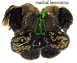
The secondary afferents leave these nuclei and
immediately cross, lining up in the ventral medulla. The new tract that they
form is called the medial lemniscus ("midline ribbon"), and it will
ascend all the way through the brainstem. Here is how it looks in the upper
medulla:

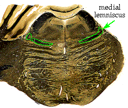
In the pons, the medial lemniscus begins to
flatten out as the pontine nuclei enlarge beneath it.

By the time we get to the midbrain, the medial
lemniscus is getting pushed way up laterally and dorsally, which will position
it to enter the thalamus.

Once in the thalamus, the secondary afferents
synapse in a thalamic nucleus called the ventrolateral posterior nucleus
(VPL). The thalamocortical afferents (from thalamus to cortex) travel up
through the internal capsule to get to primary somatosensory cortex, the
end of the pathway.
Primary somatosensory cortex is located in the
post-central gyrus, which is the fold of cortex just posterior to the central
sulcus.

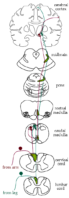
D. A diagrammatic review:
Here is the entire pathway in schematic form:

Now, think briefly about some possible
lesions. If you cut this pathway, most of the discriminative touch sensation
will be lost (not all, because some sneaks into other pathways for redundancy).
Where would the sensory loss be if you cut:
1) The left gracile fasciculus?
2) The left dorsal columns (gracile & cuneate)?
3) The right medial lemniscus, in the medulla?
4) The left internal capsule?
Answer these for yourself, with the diagram if
necessary, then scroll down.
+
+
+
+
+
+
+
+
+
+
+
+
+
+
1) The left leg and lower left trunk.
2) The left side of the body below the level
of the cut.
3) The entire left body, from the neck down.
4) The entire right body (including the face,
because the face joins the pathway in the pons, but we will get to that later).


