
Unit 5
Neuroanatomy
TOPICS COVERED
Spinal pathways
Dorsal column
Anterolateral
Somatosensory
cortex
Motor Cortex
Spinal Cord tracts
UNIT
CONTENT
Divisions of the Nervous
System:
The nervous system is comprised of central
components (the brain and the spinal cord) and peripheral components (the
sensory and motor nervous system). The sensory nervous systems receives
information from the environment and relays it to the CNS. The motor
nervous system is comprised of the somatic system and the autonomic
nervous system (ANS). The somatic nervous system innervates the muscular
system resulting in the contraction and relaxation of muscles. The ANS is
in turn divided into the sympathetic and parasympathetic nervous systems.
These two systems operate involuntarily (automatically).
Animation
1 -
Animation of the
parasympathetic and sympathetic nervous
systems
Transmission of Sensory Information within the
CNS
-
Sensory impulses are
transmitted via specialized pathways to the CNS: Dorsal column medial
lemniscal system (DCML), Anterolateral spinothalamic tract
(AST)
-
the first stage of sensory
integration and perceptual awareness occurs at the level of the
thalamus
m The
thalmas is capable of
gating out irrelevant sensory information while directing relevant
information to the cortex
-
·
sensory pathways
carrying general sensory information (e.g., touch, temperature,
proprioception) terminate in different sensory regions of the
cortex
·
Once the information
reaches the primary sensory region in the cortex we are able to recognize both the source and intensity of the sensations
·
Our final perception of
what is occurring in the environment or within our bodies is achieved once
the sensory information has been integrated and interpreted by the
associated areas of the cortex, limbic system.
The two main systems
which transmit sensory signals within the CNS:
·
The Dorsal
Lemniscal System (fine precise sensations)
·
The Anterolateral
Spinothalamic System (crude type sensations)
The type of sensations
which are characteristically conveyed in these 2 systems are tabled
below:
|
Dorsal Lemniscal
System |
Anterolateral
Spinothalamic System |
- Touch
sensations:
- High degree of
localization of stimulus.
- Fine graduations
in intensity of stimulus.
- Phasic sensations
(vibrations).
- Sensations of
movement against the skin.
- Fine
positional and pressure sensations.
|
- Thermal
sensations:
- Pain
sensations
- Crude pressure and
touch sensations
- Tickle and itch
sensations
- Sexual
sensations
|
 |
 |
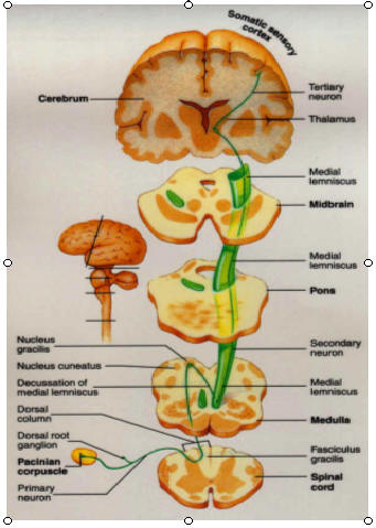
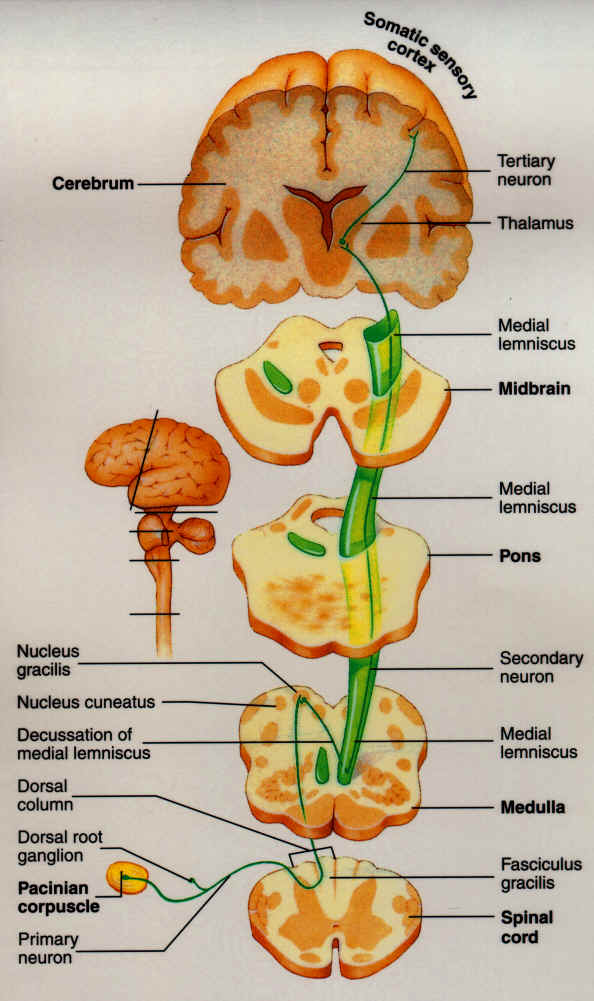
| The dorsal lemniscal system
begins with somatosensory axons entering the spinal cord via the
dorsal root and ascending in the dorsal columns ipsilaterally. The
first synapse point for this pathway is in the dorsal column nuclei
located in the medulla. The axons of neurons originating in the
dorsal column nuclei decussate (cross over), ascending via the
medial lemniscus to the contralateral ventral posterior thalamic
nucleus (VPN). Somatosensory fibers of the trigeminal nerve (CN V),
carrying information from the contralateral side of the face and
head, also synapse in the VPN. The majority of VPN neurons project
to the primary somatosensory cortex (SI), the remaining project to
the secondary somatosensory cortex (SII) of the posterior parietal
lobe. |
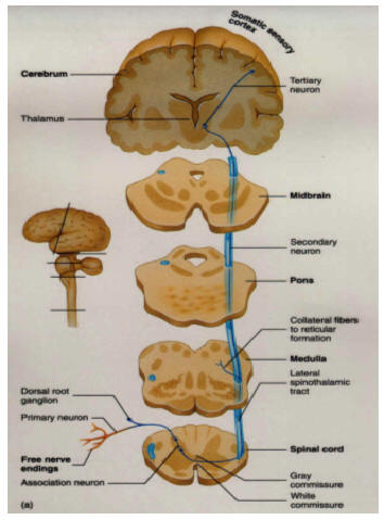
The anterolateral system
begins with somatosensory axons entering the spinal cord via the
dorsal root and synapsing upon entry. The majority of these
2nd-order axons decussate, and ascend to the brain via the
anterolateral portion of the spinal cord white matter. This
ascending system is composed of three separate tracts, the
spinothalamic tract, the spinoreticular tract, and the spinotectal
tract. The spinothalamic tract projects to the ventral posterior
nucleus of the thalamus. This tract is involved in the perception of
touch, temperature, and sharp pain. The spinoreticular tract
projects to the brain stem reticular formation on its way to the
parafasicular nucleus and intralaminar nucleus of the thalamus. This
pathway seems to be selectively involved in the perception of deep,
chronic pain. The spinotectal tract projects to the tectum of
midbrain. This tract is likely involved in some aspect of pain
perception. The tracts of the anterolateral system project to both
the primary and secondary somatosensory cortex, and to more
posterior locations within the parietal
lobe |
Somatosensory
pathways -
required reading
Neuropics
- Highly recommended – Awesome site for pathway and neuroanatomy
images
Required
QUIZ 1
UNIT
5
Please take in : www.uh.edu/webct
You will have 31 minutes to complete the Required Quiz - use your
time wisely!
Cortex
A little history
- thanks to: A Science Odyssey
| |
What would you do if you were a
doctor and had patients who were missing pieces of their skulls?
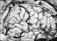
If you were Eduard Hitzig, a German doctor working at a military
hospital in the 1860s, you'd conduct some experiments. Hitzig,
working on patients who had pieces of their skulls blown away in
battle, stimulated exposed brains with wires connected to a
battery. By doing so, he discovered that weak electric shocks,
when applied to areas at the back of the brain, caused the
patients' eyes to move.
Later, around 1870, Hitzig teamed up with another doctor,
Gustav Fritsch. Setting up a makeshift lab in Fritsch's house,
the two stimulated the brains of live dogs. They found that not
only could they cause crude movements of the dogs' bodies, but
that specific areas of the brain controlled specific movements.
Soon after, John Hughlings Jackson, an English scientist,
took the work of Fritsch and Hitzig further. Based on his
observations of his wife's epilectic seizures, Jackson came up
with a more-detailed theory of how the brain controls muscles.
He knew that every one of her seizures followed the same
pattern: It would start at one of her hands, move to her wrist,
then her shoulder, then her face. It would finally affect the
leg on the same side of her body, then stop.
Jackson believed that the seizures were electrical discharges
within the brain. The discharges started at one point and
radiated out from that point. This suggested that the brain was
divided into different sections, and that each section
controlled the motor function (or movement) of a different part
of the body. And since the pattern never varied, the way the
brain is organized must also be set.
|
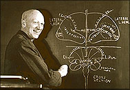
Wilder Penfield, a pioneering brain surgeon, mapped
the motor cortex using mild electric current.
Wilder Penfield took the next exploratory voyage into
the brain starting in the 1940s. While operating on
epileptic patients, Penfield applied electric currents
to the surface of patients' brains in order to find
problem areas. Since the patients were awake during the
operations, they could tell Penfield what they were
experiencing. Probing some areas triggered whole memory
sequences. For one patient, Penfield triggered a
familiar song that sounded so clear, the patient thought
it was being played in the operating room.
During these operations, Penfield watched for any
movement of the patients' bodies. From this information,
he was able to map the motor cortex, the very part of
the brain you can map in this feature's activity. |
|
Cerebral Representation of
Sensory and Motor Information (Cortex)
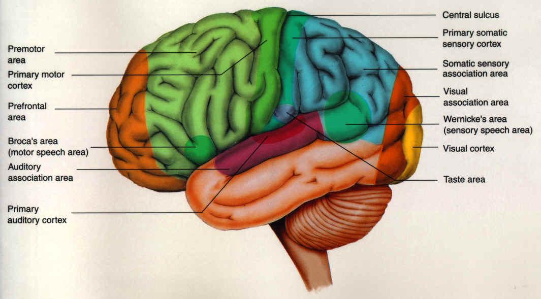
The cerebral surface is divided
into a number of groves (a sulcus) and elevations (a gyrus). A prominent
medial lateral grove is called the Central Sulcus (grove). Anterior to
this grove on the precentral gyrus resides the motor cortex. Posterior to
this grove, on the postcentral gyrus, resides the somatosensory cortex.
The motor cortex and
somatosensory cortex have a representative mapping of regions of the body.
Notice the disproportionate cortex area given to body parts used in fine
movement, such at the hands and lips for both motor and somatosensory
cortex.
Motor
Cortex I - required reading
Motor
Cortex II - required reading
Motor Cortex III
- required reading -
incredible site for entire brain - note that like the cortex, this site
has multiple layers.
Quiz questions for
the upcoming quiz will be focused on the cortex or spinal motor tracts.
Homunculus
- let this website load and have some fun learning
Spinal
motor tracts - required reading
Good
supplement - your
call
For those wanting to go the extra mile
Animation
2 -
highly
recommended – awesome site for
images, MRI scans and movies - *don't spend all day in here it would be easy to.
The Brain and Neurosyllabus sites are of most relevance to
this class. Note - it takes a little bit of time for the movies to
download so be patient, however, they really help you visualize your
neuroanatomy.
Self
Quiz -
this quiz is not graded
There are two Quizzes at the bottom of this
page - "Jigsaw Puzzle" and "Who Wants to be an Anatomist?" You will need
to download Java
Web to complete the quizzes.
You will have 31 minutes to
complete the Required Quiz - use your time wisely!
|