|
Unit 3 Skeletal Muscle and Motor Units
TOPICS COVERED
Basic structure Neuromuscular synapse General mechanism of contraction Motor units Fast and slow motor units Henneman principle Functional Role of different motor units UNIT CONTENT There are three types of muscles in the human body:
Skeletal muscle comprises the largest single organ of the body. It is highly compartmentalized, and we often think of each compartment as a separate entity (such as the biceps muscle). Each of these individual muscles is composed of single cells or fibers embedded in a matrix of collagen. At either end of the muscle belly, this matrix becomes the tendon that connects the muscle to bone. The more fibers a muscle has, the more powerful the muscle is. The quadriceps has a huge number of fibers, so it is one of the most powerful muscles in the body. Muscle cells contain most of the structures common to all cells. Each cell is enclosed by a cell membrane or plasmalemma; they contain mitochondria for the oxidative metabolism of nutrients; and all the machinery necessary for protein synthesis. Skeletal muscle fibers are multinucleated (having several nuclei) and can be as much as two centimeters long. The fibers of the quadriceps can not simply contract by themselves. Skeletal muscle has a number of components vital for proper function. Every skeletal muscle is supplied with at least one nerve – the motorneuron, one artery, and one vein. This is to ensure that every muscle can receive incoming nerve impulses while being fueled by fresh blood from the arteries. The veins are needed to export deoxygenated blood containing metabolic wastes and other byproducts. We keep talking about these "fibers" and how they contract when you flex your muscle, but what is actually happening? How is the skeletal muscle tissue able to make itself shorter to pull and move the bones? The answers to these questions lie in understanding the mechanism of muscle contraction. Very basically, muscle contraction follow this process:
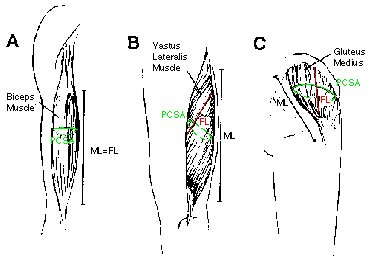
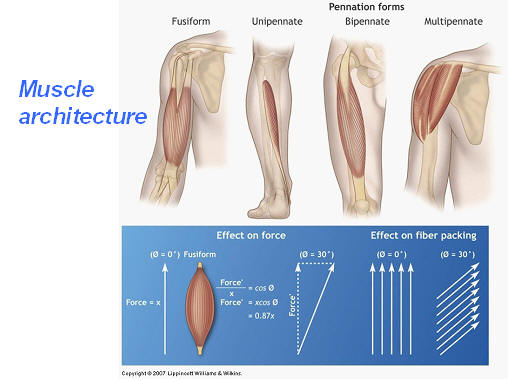
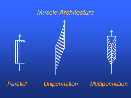
However, first we will discuss the muscle fiber arrangement, with is referred to as the muscle architecture. Skeletal Muscle Architecture Skeletal muscle is not only highly organized to function at the microscopic level, the arrangement of the muscle fibers at the macroscopic level also demonstrates a striking degree of organization. Skeletal muscle architecture is defined as "the arrangement of muscle fibers relative to the axis of force generation." The functional properties of a whole muscle depend strongly on its architecture. The various types of arrangement are as numerous as the muscles themselves, but for convenience we often refer to three types of fiber architecture. Examples of Muscle Architecture: Muscles with fibers that extend parallel to the muscle force-generating axis are termed parallel or longitudinally arranged muscles (Left). While the fibers extend parallel to the force-generating axis, they never extend the entire muscle length. Muscles with fibers that are oriented at a single angle relative to the force generating axis are termed unipennate muscles (Middle). The angle between the fiber and the force-generating axis generally varies from 0o to 30o. Most muscles fall into the final and most general category, multipennate muscles--muscles composed of fibers that are oriented at several angles relative to the axis of force generation (Right). As we will discuss, an understanding of muscle architecture is critical to understanding the functional properties of different sized muscles. A special thanks to Dr. Fred W. Kolkhorst from San Diego State University, USA for the next several figures.
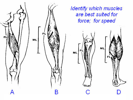
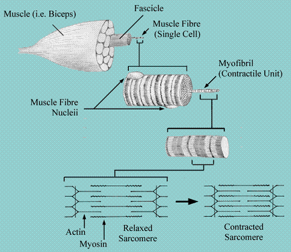
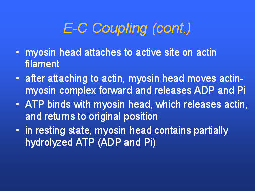
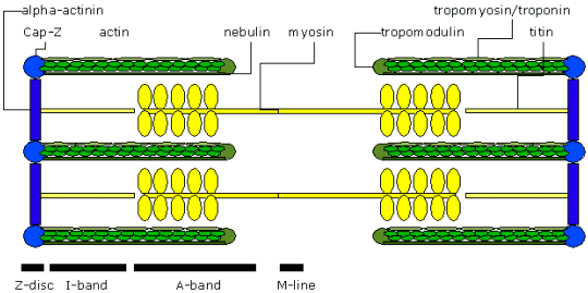
Effect of Muscle Architecture on Muscle Function The functional effect of muscle architecture can be simply stated as: muscle force is proportional to physiologic cross-sectional area (PCSA), and muscle velocity is proportional to muscle fiber length. PCSA is the sum of the areas of each fiber in the muscle. It may be apparent, based on the brief discussion of architecture presented above, that neither fiber length nor PCSA can easily be deduced based on gross muscle inspection. However, after determining architectural properties, it is possible to understand how much force the muscle generates and how fast it contracts (or how far it contracts). The principal force generating components are actin and myosin molecules. These myofilaments are arranged in interdigitating matrices capable of sliding across each other. To produce force, crossbridges from the myosin filaments associate with the actin filament, then rotate slightly to pull the filaments across each other (much like the oars of a rowboat pull the across the water). But to get to the actin and myosin (contractile fibers), let’s see the progression from muscle to myofiber to myofibril to sarcomere. The diagram below shows the organization of a typical muscle (i.e. the biceps) from a gross anatomical view, down to the microscopic anatomy of the individual units of muscle contraction. Note the organization of the muscle: contractile protein filaments are bundled into myofibrils; bundles of myofibrils are contained in each individual muscle fibre (cell); muscle fibres are bundled to form fascicles and the fascicles are bundled to form the muscle organ. The light and dark striations seen with the light microscope are caused by the very orderly arrangement of actin and myosin filaments within each myofibril and the orderly arrangement of the myofibrils within the muscle cell (fibre).
Animation
- Physiology
of muscle
Animation
– More physiology of muscle structure
This slide is a cross-sectional slice of human skeletal muscle (X40) with shows muscle fibers surrounded by connective tissue.
The myofibril itself is structured in a really amazing way that
explains how the muscle functions. The structure of the myofibril also
explains a muscle’s flexibility and its ability to contract and relax. Each
myofibril tubule is divided up into tiny sections called sarcomeres.
The sarcomere is the functional unit of muscle
tissue. This is where all the action takes place. Each one of these sarcomeres is kind of like the playing field for a huge
linked game of tug-‘o-war. The boundaries of this field are called Z lines.
You can remember this because it is the "zipper" that connects each
sarcomere. It does not actually stand for
"zipper", but it is a convenient way of remembering what the Z line
is. Like any good tug- ‘o-war, there are two major teams. The first team is a
bunch of skinny proteins called actin. The other
team is a group of burly thick proteins called myosin. This game of tug-
‘o-war is a little bit different than normal because the two teams are not
tugging on the same rope. Rather, the two teams are the ropes. Also, the
ropes are not tied together. Instead they are lined up next to each other.
The myosin proteins have little heads sticking out of the protein rope. The
tug-‘o-war happens when the myosin heads bite onto the thin actin filaments and pull the actin
in the other direction. To understand the actual mechanism of this
contraction, you must be familiar with all the boundaries and field markings
on the sarcomere.
Animation
- Sarcomere shortening
Thin filament
A special thanks to Dr. Fred W. Kolkhorst from San Diego State University, USA for the slide below.
Very cool video on actin-myosin interaction – thanks to Matthew L. Walker, Stan A. Burgess, James R. Sellers, Fei Wang, John A. Hammer III, John Trinick & Peter J. Knight. Two-headed Binding of a Processive Myosin to F-actin. Nature, 405, 804-807 (2000). PS - Be sure to click this link from the actin-myosin site above
Neat animation - follow the instructions regarding speed.
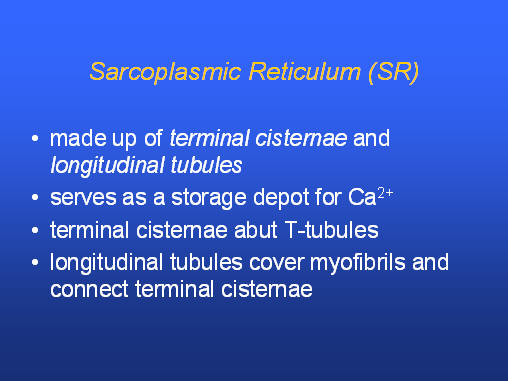
Sarcoplasmic reticulum (SR) (skeletal muscle only)
Much
of what we currently know about muscle contraction was originally reported
simultaneously by H.E. Huxley and A.F. Huxley (not related) in the 1950s and
is referred to as Huxleys' sliding filament
mechanism.
Animation
– a little actin-myosin
Animation
– sliding filament theory Animation – more sliding filament and muscle structure
Muscle
action potential
Cross-bridge cycling
Entire cycle takes 50 ms, although the myosin head is attached only 2 ms. A single myosin head will produce 3-4 pN. Animation – very good site
1st Required QUIZ UNIT
3 Please
take in : www.uh.edu/webct
You will have 36 minutes to complete the Required Quiz - use your time wisely!
Skeletal Muscle Fiber Types Not all muscle fibers within a muscle are the same; each muscle is a mixture of 3 general fiber types. Different muscles have different ratios of fiber types. Muscle fibres are usually classified based on histochemical criteria staining for the ATPase as well as metabolic capacities that reflect the activities of the glycolytic or oxidative characteristics Type I- also previously known as slow twitch, fatigue-resistant. These fibers are only aerobic and contain an abundance of mitochondria and oxidative exnzymes. The m-ATPasa enzyme activity is basic labile. These are the muscle fibers used for continuous low intensity work. Type II has three subgroups: IIa- Fast Oxidative Glycolytic- These fibers are also aerobic, and are fatigue resistant. They contain some mitochondria, oxidative and glycolytic enzymes, and the m-ATPase enzyme activity is acids labile. IIb- Fast Glycolytic- These fibers are anaerobic and easily fatigable. They contain no mitochondria and only contain gycolytic enzymes. Their only source of energy is glucose. They produce an abundance of lactic acid. Type IIb m-ATPase enzyme activity is intermediate acid stabile. They are involved in high intensity activities. IIc- Indeterminate- may become type IIa or IIb depending on the type of training. Type IIc m-ATPase enzyme activity is acid/basic stabile. Differences between fiber types ·
nerve conduction velocity ·
diameter ·
maximal tension (type II greater than type I) ·
maximal contractile speed (type II faster than type I) ·
oxidative (aerobic) capacity (type I) ·
glycolytic (anaerobic) capacity (type II) ·
fatigability
At any given velocity of movement, the amount of force produced depends on the fiber type. During a dynamic contraction, when the fiber is either shortening or lengthening, a fast-twitch (FT) fiber produces more force than a slow-twitch (ST) fiber (Fitts & Widrick, 1996). Under isometric conditions, during which the length of the muscle does not change while it is contracting, ST fibers produce exactly the same amount of force as FT fibers. The difference in force is only observed during dynamic contractions. At any given velocity, the force produced by the muscle increases with the percentage of FT fibers and, conversely, at any given force output, the velocity increases with the percentage of FT fibers.
Table of fiber type characteristics
There is great variability in the percentage of fiber
types among athletes. For example, it is well known that endurance athletes
have a greater proportion of slow-twitch fibers, while sprinters and jumpers
have more fast-twitch fibers (Costill, et al., 1976;
Ricoy, et al., 1998). The greater percentage of FT
fibers in sprinters enables them to produce greater muscle force and power
than their ST -fibered counterparts (Fitts & Widrick, 1996). Differences in muscle fiber com- position
among athletes have raised the question of whether muscle structure is an
acquired trait or is genetically determined. Studies performed on identical
twins have shown that muscle fiber composition is very much genetically
determined (Komi & Karlsson,
1979), however there is evidence that both the structure and metabolic
capacity of individual muscle fibers can adapt specifically to different
types of training. Can fiber types be changed by training?
NEUROMUSCULAR JUNCTION
Animation – muscle activation Link to
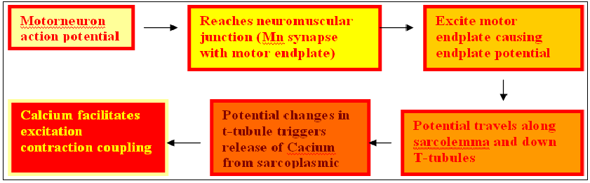
neuromuscular junction figure
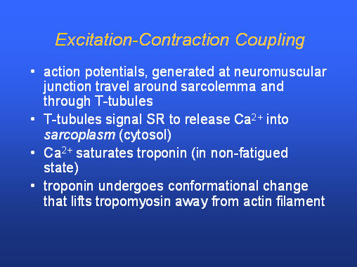
1. nerve impulse leads to 2. ACh release 3. ACh binds to ACh receptors 4. Na+ influx ---> action potential down t-tubules, leading to excitation-contraction coupling.
Animation - active neuromuscular junction
Practice Quiz – not graded but good fun Muscle fibers are innervated by neurons whose cell bodies are located in spinal cord. The nerve fibers, or axons, of these motor neurons leave the spinal cord and are distributed to the motor nerves. Each motor axon branches several times and innervates many muscle fibers. The combination of a single motor neuron and all the muscle fibers it innervates is called a motor unit. Although the muscle fibers of a given motor unit tend to be located near one another, motor units have overlapping territories. In response to an action potential from the neuron, a muscle fiber depolarizes as the signal propagates along its surface and the fiber twitches (contracts). This depolarization generates an electric field in the vicinity of the muscle fibers which can be detected by a skin surface electrode located near this field, or by a quadrifillar electrode inserted in the muscle. The resulting signal is called the muscle fiber action potential. The combination of the muscle fiber action potentials from all the muscle fibers of a single motor unit is the motor unit action potential (MUAP). All of the muscle fibers in a motor unit are fired each time a motor unit fires. The repetitive firing of a motor unit creates a train of impulses known as the motor unit action potential train (MUAPT). The summation of electrical activity created by each active motor unit is the myoelectrical signal (ME) (4). To sustain muscle contraction, the motor units must be repeatedly activated (2). As the firing rates of motor units active in a contraction increase, the twitches associated with each firing will eventually fuse to yield large forces. Link to Muscle Architecture figure
The force of contraction is controlled with the number of motor units recruited firing rate and also the type of motor units recruited. As a general rule,
motor units are recruited in order of their size. When the muscle is
activated initially, the first motor units to fire are small in size and weak
in the degree of tension they can generate. Starting with the smallest motor
units, progressively larger units are recruited with increasing strength of
muscle contraction. The result is an orderly addition of sequentially larger
and stronger motor units resulting in a smooth increase in muscle strength. The recruitment sequence is thought to begin with type I motor units, to progress to type II units that first include type IIa, and to end with type IIb units, which are active only at relatively high force output.
2nd Required QUIZ UNIT 3 Please take in : www.uh.edu/webct You will have 16 minutes to complete the Required Quiz - use your time wisely! |