|
Unit 7 Reflexes
TOPICS COVERED
Reflexes Reflex arc Stretch reflex H-reflex Golgi tendon reflex Withdraw reflex
UNIT CONTENT
What is a reflex?
reflex -- a simple, relatively stereotyped action caused by a specific stimulus Reflexes are rapid, involuntary responses to stimuli which are mediated over simple nerve pathways called reflex arcs. Involuntary reflexes are very fast, traveling in milliseconds. The fastest impulses can reach 320 miles per hour.
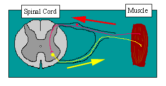
Reflex arcs have five essential components:
1. The receptor at the end of a sensory neuron reacts to a stimulus. 2. The sensory neuron conducts nerve impulses along an afferent pathway towards the CNS. 3. The integration center consists of one or more synapses in the CNS. 4. A motor neuron conducts a nerve impulse along an efferent pathway from the integration center to an effector. 5. An effector responds to the efferent impulses by contracting (if the effector is a muscle fiber) or secreting a product (if the effector is a gland).
Reflexes can be categorized
as either autonomic or somatic. Autonomic reflexes are not subject to
conscious control, are mediated by the autonomic division of the nervous
system, and usually involve the activation of smooth muscle, cardiac
muscle, and glands. Somatic reflexes involve stimulation of skeletal
muscles by the somatic division of the nervous system.
The receptors described in Unit 4 are all involved in various ‘reflexes’.
Tonicity of Skeletal Muscle
Tone within skeletal muscle is controlled via a receptor called the MUSCLE SPINDLE. Therefore, to understand the control of tone it is imperative to understand function of the muscle spindle.
The functional value of reflexes
The Stretch Reflex
As briefly described above the muscle spindle plays an integral role in the stretch reflex. In brief: As a muscle lengthens the MS is stretched. Impulses are conducted towards the CNS (spinal cord) where the afferent fiber divides into several colateral fibers. One of these colateral fibers stimulates the homonymous muscle (same muscle that was stretched) causing it to contract which in turn relieves the stretch stimulus to the muscle spindle. Simultaneously, another afferent collateral synapses with an inhibitory interneuron (Renshaw cell secreting GABA) which in turn synapses on the neuron innervating the antagonistic muscle (opposing muscle to which was stretched).
Highly recommended
–
See it in action – animation of tendon tap
Innervation of the Muscle Spindle The nerve fibers attached to the muscle spindle either conduct impulses from the Spindle to the CNS (afferent/sensory fibers) or from the CNS towards the muscle (efferent/motor fibers).
Afferents:
Type 1a fibers: 17 microns in diameter, conduct impulses at 100m/s. secondary endings (flower spray endings) Type II Fibers: 8 Microns in diameter
Efferents:
Alpha motorneuron 120 m/s). (from the CNS) Gamma motor fibers
Stretch reflex Ia primaries afferents have powerful excitatory effect on a motoneurones of same muscle and synergists in adjacent spinal segments. May be monosynaptic or polysynaptic.
Reciprocal inhibition: Ia also inhibit a motoneurones of antagonistic muscles via inhibitory interneurone and corresponding contralateral muscles. Ia afferents also have a weak polysynaptic excitatory action on dynamic and static gamma motoneurones.
Group II afferents from spindle secondaries also excite autogenic alpha motoneurones via mono & polysynaptic paths. Monosynaptic component involves about 50% of the motoneurones that are excited by Ia gamma motoneurones. highly responsive to electical stimulaton of group II afferents (but not clear how much of this group II input is purely spindle in origin). Classical stretch reflex 'the capacity of a muscle to resist extension' is sum of these spindle projections to muscle. The monosynaptic Ia component is responsible for the 'tendon jerk' . The 'tonic stretch reflex' is mainly disynaptic or polysynaptic
2) How does the muscle spindle contribute to the automatic regulation of muscle length? - a) the stretch reflex is an example of muscle spindle sensory and motor function b) stimulated muscle spindle sends message to the spinal cord, triggering alpha motoneuron firing, which, in turn, causes contraction of the lengthened muscle
Alpha motor neurons and motor units contribute to muscle contraction
Cerebellar 'awareness':
After Muscle Spindle stimulation (stretch) and the afferent fiber enters into the spinal it divides into several colaterals. Some of these colaterals synapse on the cell bodies of neurons which ascend to the cerebellum (anterior and posterior spinocerebellar tracts). Thus, at all times the cerebellum is aware of the state of stretch in muscles, in other words the TONE of muscles.
Coactivation of Gamma efferents Whenever a motor command descends from the motor cortex and synapses on neural cell bodies which innervate muscles, collaterals from these descending fibers also synapse on the corresponding cell bodies (gamma efferents) which innervates the ends of the intrafusal muscle fibers. This is important so that as the extrafusal muscle fibers contract and shorten, the intrafusal also shorten and remain taunt. This enable the MS to always respond to stretch even immediately after contraction of a muscle. In other words the coactivation of gamma efferents avoids 'silent periods' which would occur if the intrafusal muscle fibers did not contract simultaneously with the extrafusal muscle fibers.
Thus with gamma drive, the spindle is ready to respond to unexpected perturbation The spindle activity generates a reflex response which compensates for the perturbation.
How to increase the stretch reflex
1.
Jendrassik's maneuver: 2. Gripping an object.
HOW? Tendon jerk is reinforced by clenching fists or jaw as the Gamma pathway is centrally facilitated rendering spindle more sensitive to stretch. H-reflexHoffmann Reflex (H-Reflex) technique.
The H-reflex and F-wave
H-Reflex
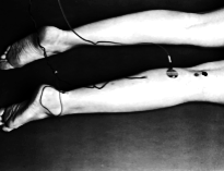
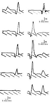
The H-reflex is the electrical equivalent of the monosynaptic stretch reflex and is normally obtained in only a few muscles. It is elicited by selectively stimulating the Ia fibers of the posterior tibial or median nerve. Such stimulation can be accomplished by using slow (less than 1 pulse/second), long-duration (0.5-1 ms) stimuli with gradually increasing stimulation strength. The stimulus travels along the Ia fibers, through the dorsal root ganglion, and is transmitted across the central synapse to the anterior horn cell which fires it down along the alpha motor axon to the muscle. The result is a motor response, usually between 0.5 and 5 mv in amplitude, occurring at low stimulation strength, either before any direct motor response (M) is seen or with a small M preceding it. Understandably, the latency of this reflex is much longer than that of the M response, and a sweep of 5-10 ms/division is necessary to see it.
The H-reflex can normally be seen in many muscles but is easily obtained in the soleus muscle (with posterior tibial nerve stimulation at the popliteal fossa), the flexor carpi radialis muscle (with median nerve stimulation at the elbow), and the quadriceps (with femoral nerve stimulation).
Typically, it is first seen at low stimulation strength without any motor response preceding it. As the stimulation strength is increased, the direct motor response appears. With further increases in stimulation strengths, the M response becomes larger and the H-reflex decreases in amplitude. When the motor response becomes maximal, the H-reflex disappears and is replaced by a small late motor response, the F-wave.
H-reflex latency can be determined easily from charts, according to height and sex or from published normal values. Whatever these values however, the best normal value in localized processes is the patient's asymptomatic limb. If no facilitation maneuvers are performed, the difference in latency between both sides should not exceed l ms. The H-reflex is useful in the diagnosis of S1 and C7 root lesions as well as the study of proximal nerve segments in either peripheral or proximal neuropathies. Its absence or abnormal latency on one side strongly indicates disease if a local process is suspected. Much controversy remains, however, on whether its absence bilaterally in otherwise asymptomatic individuals is of any clinical significance.
F-Wave The F-wave is a long latency muscle action potential seen after supramaximal stimulation to a nerve. Although elicitable in a variety of muscles, it is best obtained in the small foot and hand muscles. It is generally accepted that the F-wave is elicited when the stimulus travels antidromically along the motor fibers and reaches the anterior horn cell at a critical time to depolarize it. The response is then fired down along the axon and causes a minimal contraction of the muscle. Unlike the H-reflex, the F-wave is always preceded by a motor response and its amplitude is rather small, usually in the range of 0.2-0.5 mv.
The F-wave is a variable response and is obtained infrequently after nerve stimulation. Commonly, several supramaximal stimuli are needed before an F-response is seen since only few stimuli reach the anterior horn cell at the appropriate time to depolarize it. With supramaximal stimulation however, depolarization of the entire nerve helps spread the stimulus to the pool of anterior horn cells thus enhancing its chances to reach a greater number of anterior horn cells at the critical time and produce an F-wave.
Because different anterior horn cells are activated at different times, the shape and latency of F-waves are different from one another. Conventionally, ten to twenty F-waves are obtained and the shortest latency F-wave among them is used.
The normal values can be determined from charts or published data and, in unilateral lesions, the best normal values remain those of the patient's asymptomatic limb. The difference between both sides' shortest latencies should not exceed l ms. The data obtained from the F-wave have been used in many different ways to determine proximal or distal pathology. Those include the F-wave chronodispersion or difference in latency between the F-wave with the shortest and that with the longest latency, and the F-wave ratio. We find the F-wave ratio very useful in routine clinical work. It is obtained by dividing the conduction time of the proximal nerve segment by that of the distal nerve segment and is performed as follows: Obtain the F-wave latency from proximal (F prox) stimulation (knee or elbow). Obtain the motor response likewise from proximal stimulation (M prox). Then determine the latency of the proximal nerve segment by this equation:
Proximal latency = (Fprox - Mprox - 1 ms) / 2
where l ms is the estimated delay encountered by the stimulus at the anterior horn cell. The latency of the distal segment is none other than the motor response latency obtained from proximal stimulation (M prox). The F-ratio is then obtained by dividing the proximal latency by the distal latency:
F-ratio = (Fprox - Mprox - 1 ms) / 2 x Mprox
1st Required QUIZ Unit 7
Please
take in:
www.uh.edu/webct
You will have 22 minutes to complete the Required Quiz - use your time wisely!
Golgi tendon reflex
This reflex regulates tension e.g. When attempting to maintain a steady grip on a cup
Inhibition of the alpha motor neurons causes muscle relaxation, relieving the tension in the muscle.
The Withdrawal reflex (flexor/crossed extensor reflex) - its action is to withdraw a limb from a noxious stimulus.
For example, If you step on a sharp object you stimulate pain and cutaneous receptors from skin and muscle. This elicits both excitation of synergistic muscles and inhibition of antagonist muscles, say, in your legs; as well as contracting extensors and inhibiting flexors on the opposite side to maintain posture and balance.
Tonic Vibration Reflex and Vibration Training
Tonic Vibration reflex - in Latash - pages 76-77
Tonic vibration reflex - vibration can drive primary afferents - driving is when an action potential is induced in response to every cycle of the stimulus. When a muscle is vibrated it produces a tonic muscle contraction known as a Tonic Vibration Reflex (TVR)
The responses to muscle
vibration are unique for a variety of reasons: 2) monosynaptic reflexes are inhibited during TRV - monosynaptic inputs are inhibited presynaptically but polysynaptic inputs remain excitatory - hence tonic muscle contraction 3) muscles not subject to vibration display reflex responses (responses can be intersegmental) 4) vibration produces illusions
Good starting point - mandatory - material from this paper is fair game for your quiz
Mandatory paper 1 - (i.e. material from this paper is fair game for your quiz)
Mandatory paper 2 - (i.e. material from this paper is fair game for your quiz)
Galileo Vibration training system
Power Plate site - see research abstracts
2nd Required QUIZ Unit 7
Please
take in:
www.uh.edu/webct You will have 12 minutes to complete the Required Quiz - you know the drill :) |