|
Unit 6 Basal Ganglia and Cerebellum
TOPICS COVERED Basal Ganglia anatomyBasal ganglia pathways Basal ganglia function Basal ganglia disorders
Cerebellum anatomy Cerebellum pathways Cerebellum function Cerebellum disorders
UNIT CONTENT
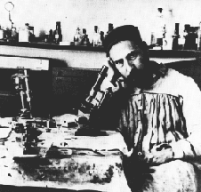
History
Thanks to: The Institute for Adaptive and Neural Computation (ANC) is part of the School of Informatics at the University of Edinburgh.
The basal ganglia - required readingThe term "basal ganglia" refers to a loosely grouped collection of large subcortical nuclear (gray) masses derived from the telencephalon and located deep within each cerebral hemisphere. The nuclear components include the claustrum, amygdaloid nuclear complex, nucleus accumbens, and the corpus striatum. The term is now used to refer more specifically to the corpus striatum and two additionally related nuclei, the subthalamic nucleus and the substantia nigra; derivatives of the diencephalon and mesencephalon, respectively. Although not part of the telencephalic basal ganglia these nuclei are reciprocally connected with and functionally related to the corpus striatum. The focus of this tutorial will be entirely on the corpus striatum and those structures functionally related to it. We know from our understanding of their organizaton, connections, and deficits that ensue from their pathology that the corpus striatum and related nuclei play an important role in motor control. Interestingly, the component nuclei themselves have no direct projections to spinal cord levels. They influence motor activity by interacting with areas of the cerebral cortex that give rise to descending motor pathways through a number of feedback loops. The corpus striatum, related nuclei, and connections are sometimes described as forming the "extrapyramidal motor system." This is now an antiquated concept but was once conceived to represent a complete and independent motor system distinct from the phylogenetically newer corticospinal or "pyramidal motor system". Sweet,sweet pictures - take a look, I suspect you will find them useful.
Thanks to:
Basal ganglia II - required reading Special thanks to the Karolinska Institutet © for the material immediately below. Basal ganglia sagittal view depiction
Basal ganglia dorsal plane view
MRI of the caudate nucleus
Disorders of the basal ganglia Wilson's disease - required reading Huntington disease - required reading Tourette's syndrome -required reading
1st Required QUIZ UNIT 6 Please take in : www.uh.edu/webct You will have 22 minutes to complete the Required Quiz - use your time wisely!
Parkinson's disease - required reading - be sure to read the entire side (all segments listed in the left column)
2nd Required QUIZ UNIT 6 Please take in : www.uh.edu/webct You will have 8 minutes to complete the Required Quiz - use your time wisely!
The Cerebellum - required reading · Function: To enable smooth coordinated movement · plays an important role in the maintenance of posture and equilibrium. · Sends fibers via the cerebellar-spinal tract to modulate the activity of the alpha as well as the gamma motor neurons (Lower motor neurons). o The cerebellum receives information from a wide array of senses: o Pressure and touch o Positional receptors: Spindle and Golgi tendon organs o Eyes and ears o A copy of the command signal is sent from the cerebral cortex to the cerebellum, where it acts as a comparator.
Cerebellum II - required reading
WWAMI MEDICAL EDUCATION PROGRAM Washington-Wyoming-Alaska-Montana-Idaho
Special thanks to the Karolinska Institutet © for the pictures immediately below.
Basal ganglia sagittal view depiction
MRI of the cerebellum
Detailed MRI for those interested in the technology - NOT required reading
Cerebellum III - required reading - a special thanks to New Horizons for Learning for this material.
Cerebellum IV - required reading - a special thanks to the Society for Neuroscience for this material Clinical deficits associated with cerebellar lesions: - REQUIRED READING - just know basic symptoms
Just a great listing of all sorts of "neuro" stuff. - Not required but interesting (some links are dead) 2nd Required QUIZ UNIT 6 Please take in : www.uh.edu/webct You will have 25 minutes to complete the Required Quiz - use your time wisely! |
 At the beginning of the 20th century there were serious attempts
to provide detailed comparative descriptions of the corpus
striatum (Wilson 1914,
At the beginning of the 20th century there were serious attempts
to provide detailed comparative descriptions of the corpus
striatum (Wilson 1914,


